Will Sciatica Show On MRI? Here's What You Need to Know
This article explores how this imaging tool unveils the intricate details of spinal structures, revealing herniated discs or spinal stenosis that contribute to sciatic nerve compression.
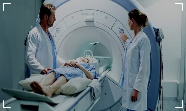
Sciatica is a term that describes the symptoms of leg pain—and possibly tingling, numbness, or weakness—that originate in the lower back and travel through the buttock and down the large sciatic nerve in the back of each leg.
In this article, we uncover:
- MRI scans and the relation between Sciatica diagnosis.
- Identifying underlying conditions.
- Help and interpret the results.
What is Sciatica and How Does it Present Itself?
Sciatica is not a medical diagnosis in itself – it is a symptom of an underlying medical condition.
It's typically characterized by a sharp pain that radiates from the lower back down to the legs, which can vary in frequency and intensity.
Some patients experience a mild ache, while others describe the pain as sharp, burning, or even electric-like.
Certain movements or positions can exacerbate the discomfort, and it may be accompanied by numbness or muscle weakness along the nerve pathway.
MRI in Diagnosing Sciatica
When it comes to diagnosing the root cause of sciatica, an MRI (Magnetic Resonance Imaging) is often the go-to imaging technique.
This is because an MRI provides detailed images of the body's soft tissues, including the spinal cord, nerve roots, and intervertebral discs.
It can reveal herniated discs, bone spurs, spinal stenosis, or tumors that may be pressing on the sciatic nerve.
However, it's important to note that while an MRI can show these conditions, it doesn't always correlate with the presence or severity of sciatica symptoms.
MRI Determining Sciatica
Many doctors use an MRI for sciatica diagnoses. Instead of radiation, MRI scans use a strong magnetic field and radio waves to capture high-definition images of your bones, tissues, and organs. It's one of the safest imaging techniques to capture herniated discs that may be causing your sciatica.
For the evaluation of sciatica pain, a lumbar spine MRI is typically recommended. This specific type of MRI focuses on imaging the lower part of the spine, including the lumbar vertebrae, discs, and surrounding structures.
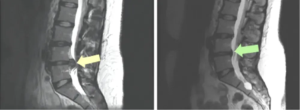
A lumbar spine MRI provides detailed images that can reveal conditions such as herniated discs, spinal stenosis, or other issues contributing to the compression or irritation of the sciatic nerve.
This targeted imaging approach allows healthcare professionals to assess the underlying causes of sciatica and formulate an appropriate treatment plan based on the findings.
Understanding MRI Technology
Magnetic Resonance Imaging (MRI) is a non-invasive diagnostic tool that uses a powerful magnetic field, radio waves, and a computer to produce detailed pictures of the inside of your body.
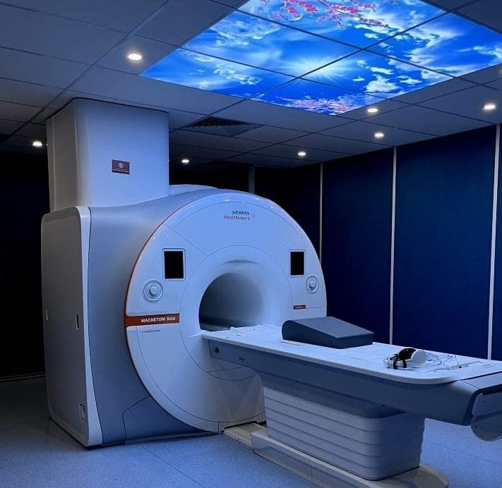
Unlike X-rays or CT scans, MRIs do not use ionizing radiation, which makes them a safer option for repeated use.
The technology is particularly adept at imaging non-bony parts or soft tissues of the body, which is why it's so useful for detecting potential causes of sciatica.
Preparing for an MRI Scan
If your doctor has recommended an MRI to investigate your sciatica, there's some preparation involved.
You'll need to remove any metal objects, as the magnetic field can attract metals and cause harm or affect the image quality.
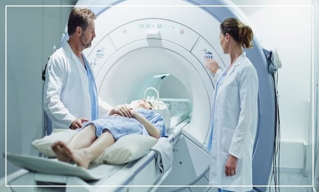
You'll also be asked about any implants, such as pacemakers or cochlear implants, which may be affected by the MRI.
It's a painless procedure, but some people may feel claustrophobic in the machine. In such cases, a sedative may be offered.
What Can an MRI Show?
An MRI can reveal a variety of conditions that may be causing sciatica. For example, a herniated disc, which occurs when the soft center of a spinal disc pushes through a crack in the tougher exterior casing, is a common cause of sciatic nerve pain.
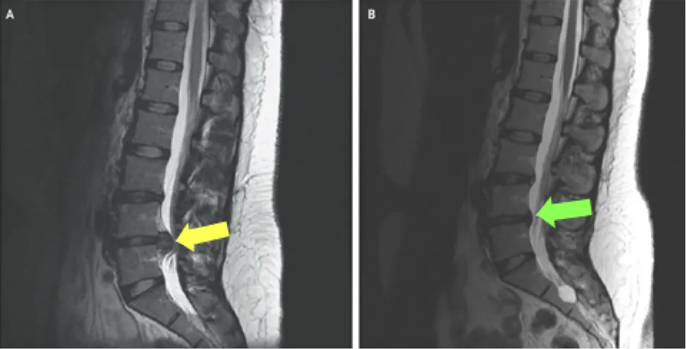
An MRI can also show spinal stenosis, a narrowing of the spinal canal that compresses the nerves. Other detectable conditions include spondylolisthesis (slipped disc), degenerative disc disease, and in rare cases, tumors or infections.
Limitations of MRI
While MRIs are incredibly useful, they have their limitations. Sometimes, an MRI can show abnormalities that may not be causing any symptoms.
Conversely, some patients with severe sciatica symptoms may have MRIs that show only minor abnormalities or even appear completely normal.
This discrepancy is why a comprehensive clinical evaluation by a healthcare professional is crucial in conjunction with imaging results.
Interpreting MRI Results
Interpreting an MRI requires expertise. Radiologists are trained to read MRI images and report their findings to your doctor.
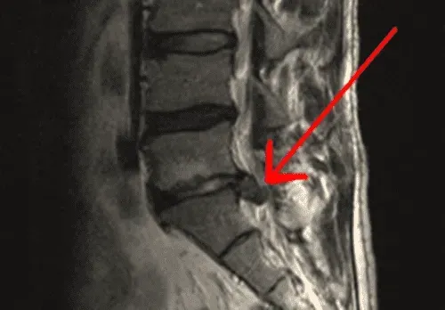
The report will detail any abnormalities found and their potential significance. Your doctor will then use this information, physical examination, and medical history to arrive at a diagnosis and develop a treatment plan.
Treatment Options
Once the MRI has identified the cause of sciatica, treatment options can be more accurately determined.
These may include physical therapy, medications, injections, or in some cases, surgery. The goal of treatment is to alleviate pain and address the underlying condition to prevent recurrence.
Summary
Sciatica can be a debilitating condition but with the help of MRI technology, the underlying causes can often be identified and treated. While an MRI can show the conditions that are likely to cause sciatica, it's important to remember that the scan is just one part of the diagnostic process. A thorough clinical evaluation is essential to ensure accurate diagnosis and effective treatment. If you're struggling with sciatica, an MRI may be a valuable step in getting to the root of the problem and finding relief.
FAQ Section
Q 1: Can Sciatica be Diagnosed Through an MRI?
A: Yes, Sciatica can often be diagnosed through an MRI. Magnetic Resonance Imaging (MRI) is a valuable diagnostic tool for identifying the underlying causes of sciatic nerve pain.
It provides detailed images of the spine, allowing healthcare professionals to visualize potential issues such as herniated discs, spinal stenosis, or other conditions compressing the sciatic nerve. However, while MRI is instrumental, a comprehensive diagnosis may also involve clinical evaluation and consideration of symptoms.
Consult with a healthcare professional for an accurate diagnosis and tailored treatment plan.
Q 2: What MRI Findings Indicate Sciatica?
A: MRI findings related to sciatica can provide valuable insights into the underlying causes of this condition. Common observations on an MRI scan that may indicate sciatica include:
- Disc Herniation:
The presence of a herniated or bulging disc, particularly in the lumbar spine (lower back), can compress the sciatic nerve, leading to pain and discomfort. - Nerve Compression:
MRI scans can reveal signs of nerve compression, such as the narrowing of the spinal canal or foraminal stenosis, contributing to sciatic nerve impingement. - Inflammation:
Inflammatory changes around the sciatic nerve or surrounding tissues may be evident, suggesting irritation and contributing to sciatica symptoms. - Disc Degeneration:
Degenerative changes in the intervertebral discs, like disc degeneration or loss of disc height, can contribute to sciatica by reducing the space available for nerve roots. - Cysts or Tumors:
The presence of cysts or tumors near the sciatic nerve pathway may exert pressure on the nerve, causing sciatica symptoms. - Spinal Stenosis:
MRI can identify spinal stenosis, a condition where the spinal canal narrows, potentially impacting the sciatic nerve and causing pain. - Soft Tissue Abnormalities:
Abnormalities in the soft tissues surrounding the sciatic nerve, such as muscle inflammation or swelling, may be visible on an MRI.
It's important to note that MRI findings are just one part of the diagnostic process. Clinical evaluation and a thorough medical history are also crucial for a comprehensive diagnosis.
If you suspect sciatica, consulting with a healthcare professional who can interpret these MRI findings in the context of your specific symptoms is essential for effective management and treatment.
Q 3: Are Other Diagnostic Tests Necessary Alongside MRI for Sciatica?
A: Yes, while MRI is a valuable tool for diagnosing sciatica, additional diagnostic tests may be necessary for a comprehensive evaluation. X-rays can help assess bone health and identify issues like spinal stenosis or fractures. Electromyography (EMG) measures muscle activity and helps pinpoint nerve compression.
Furthermore, nerve conduction studies (NCS) can evaluate how well electrical impulses travel along nerves, aiding in identifying nerve damage. These supplementary tests, along with a thorough clinical examination, provide a more holistic understanding of the underlying causes of sciatica, enabling healthcare professionals to tailor effective treatment plans.
To delve deeper into this topic, you may find our blog article on "Comprehensive Diagnostics for Sciatica: Beyond MRI" insightful. Feel free to explore the article for a detailed overview of the various diagnostic tests and their roles in assessing sciatica.
